Research | 2021-06-01
BackRecently, a research team led by Professor Changfeng Wu from the Department of Biomedical Engineering at the Southern University of Science and Technology (SUSTech) developed a series of highly bright polymer dots probes. Through the functionalization of polymer dots probes and application in expansion microscopy, fine subcellular structures with a resolution of ≈ 30 nm can be resolved on a conventional fluorescent microscope. Their research, entitled “Expansion Microscopy with Multifunctional Polymer Dots,” was published in Advanced Materials, a top journal in the field of materials science.
Super-resolution optical imaging won the 2014 Nobel Prize in Chemistry for its ability to provide resolution below the diffraction limit. The current super-resolution technology mainly contains two categories, one of which relies on patterned illumination modulation and the other one based on single-molecule positioning. Expansion microscopy uses a completely different strategy that physically enlarges the samples to allow clear distinction of adjacent molecules that are originally within the diffraction limit. This method does not rely on a complex imaging system, and nanoscale resolution can be achieved on a common confocal microscope. However, the fluorescence brightness attenuation caused by chemical quenching and density dilution during sample preparation has long been a challenge for further applications.
Changfeng Wu’s research group developed multifunctional polymer dots for application in multicolor expansion microscopy to address this issue. The fluorescence intensity of Pdots in ExM was up to 6 times higher than those achieved using commercially available Alexa dyes. The impressive brightness of the Pdots facilitated multicolor ExM, thereby enabling a variety of subcellular structures, such as mitochondria, clathrin-coated pits, and neuron synapses to be visualized on traditional fluorescent microscopes (Figure 1a-c). Furthermore, the research group combined polymer dots probes, expansion microscopy, and super-resolution optical fluctuation microscopy to achieve ultrahigh-resolution imaging of subcellular structures on a conventional wide-field microscope. As a result, this reflected the actual size of microtubules and the hollow membrane structure of mitochondria (Figure 1d-j). These findings highlight the immense potential of highly bright polymer dots for biological imaging.
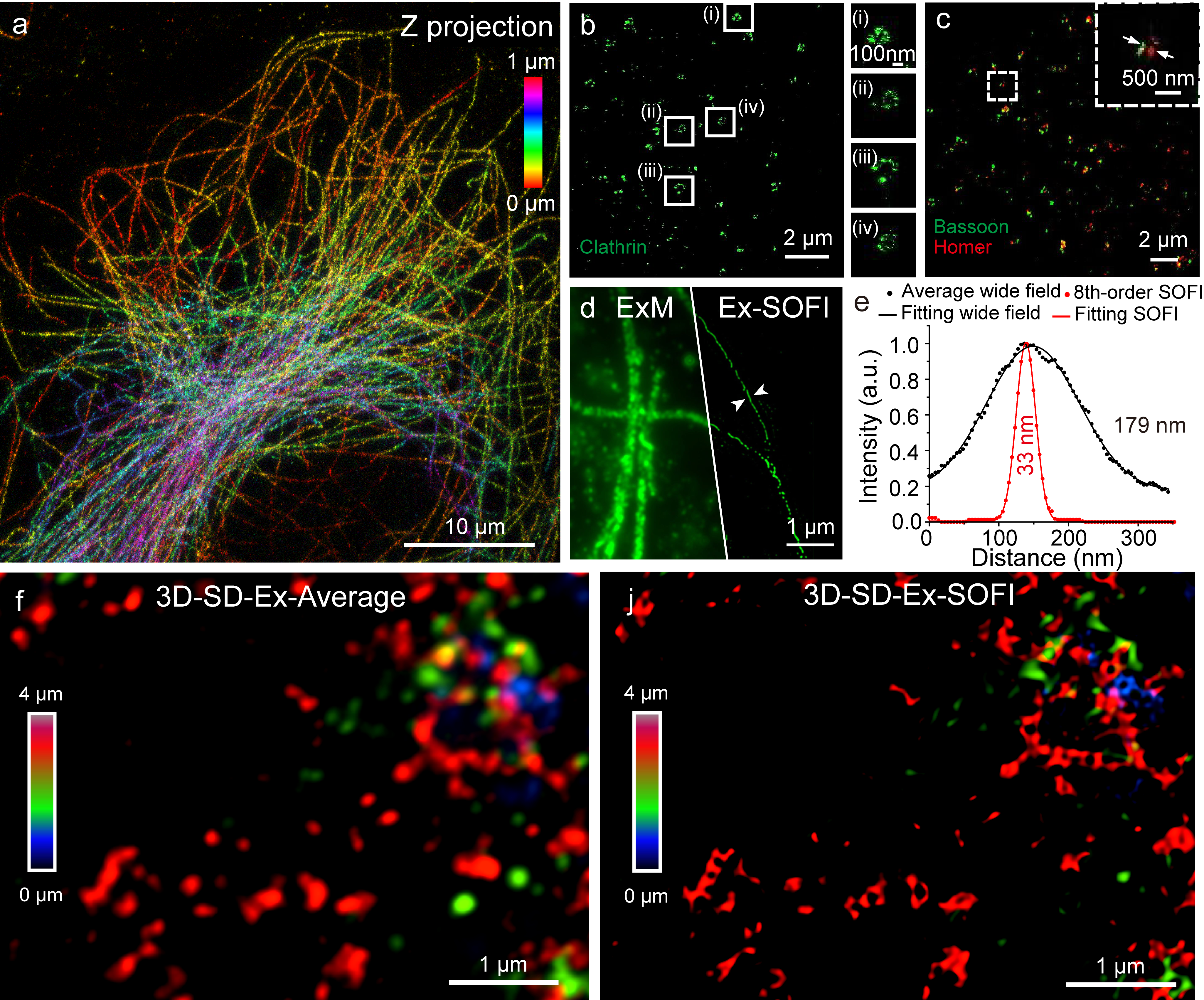
Figure 1. Three-dimensional super-resolution expansion and optical fluctuation imaging of subcellular structures
The immunofluorescence staining of samples is tedious and time-consuming. To improve the efficiency of the project, Zhihe Liu, a postdoctoral fellow in the research group, designed an automatic cell immunostaining system (Figure 2). As a result, this could replace manual work for immunofluorescence staining experiments.
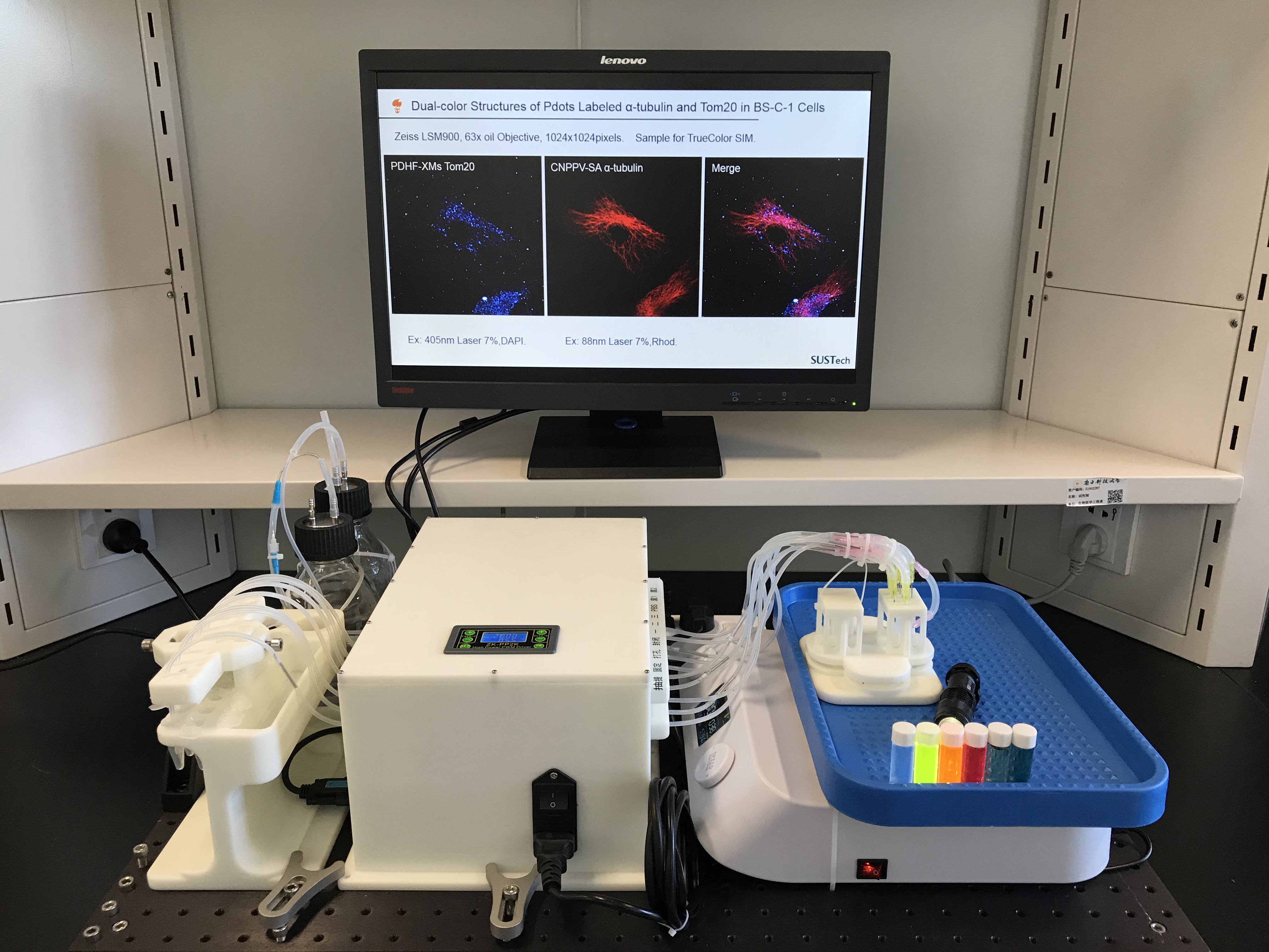
Figure 2. Automatic cell immunostaining system
Jie Liu, a doctoral student supported by the joint Ph.D. program between SUSTech and the Hong Kong Baptist University (HKBU), is the first author of this paper. SUSTech is the correspondence unit of this paper. The above research has been supported by the National Natural Science Foundation of China (NSFC), the National Key R&D Program of China, and the Shenzhen Science and Technology Innovation Commission.
Paper link: https://doi.org/10.1002/adma.202007854


Faculty Research Building 2, 1088 Xueyuan Rd, Nanshan District, Shenzhen, Guangdong 518055, China
bme@sustech.edu.cn

关注微信公众号


